Sep 16, 2016

DNA MICROARRAY PROTOCOL

i)           Set-up the following Pre-Hybridisation solution in a Coplin Jar and        incubate at 65°C during the labeling incubation period to equilibrate. 20X SSC 8.75 ml 20% SDS 0.25 ml BSA (100 mg/ml) 5.0 ml H2O to 50.0 ml

ii)            Label control and test genomic DNA as follows:- CONTROL TEST Genomic DNA ˜ 2 mg ˜ 2 mg Random Hexamers (3 mg/ml) 1 ml 1 ml H2O to 41.5 ml to 41.5 ml Heat at 95ºC for 5 minutes. Snap cool on ice and briefly centrifuge. 10X buffer 5 ml 5 ml dNTP's (5mM each dATP, dGTP & dTTP, 2mM dCTP) 1 ml 1 ml Cy-labelled dCTP 1.5 ml (Cy3) 1.5 ml (Cy5) Klenow fragment (10U/ml) 1 ml 1 ml Incubate at 37°C for 90 minutes.

iii)          Incubate the microarray slide(s) in the Pre-Hybridisation solution for 20 minutes at 65°C, beginning just before the end of the labelling reactions incubation time at 37°C.

iv)          Combine the control and test reactions and purify using the Qiagen MinElute PCR Purification kit, using a two step wash stage using 500 ml then 250 ml volumes of Buffer PE and eluting the labeled cDNA from the MinElute column with 14 ml H2O. The columns retain approximately 1 ml, so the final eluted volume will be 13 ml.

v)           Rinse the pre-hybridised microarray slides in H2O for 1 minute, then in isopropanol for 1 minute. Spin at 1500 rpm for 5 minutes to dry slides. Keep in covered slide box. 1 NICK DORRELL - LAST UPDATE FEBRUARY 2004

vi)          Prepare the Hybridisation solution as follows: - Sample 13 ml H2O 26 ml 20X SSC 12 ml 2% SDS 9 ml Heat at 95ºC for 2 minutes. Allow to cool slowly at room temperature and centrifuge for 30 seconds. Add 2 x 20 ml H2O to the corners of the hybridisation chamber. Place a slide into the chamber. Place a LifterSlip™ glass coverslip (22 mm x 25 mm) over the array section on the slide using tweezers. Pipette the Hybridisation solution onto the slide at the top of the coverslip. Seal the chamber and incubate in a water bath at 65°C overnight.

vii)         Prepare Wash solutions as follows: - Wash A (1X SSC 0.5% SDS) Wash B (0.06X SSC) 20X SSC 20 ml 2.4 ml 20% SDS 1 ml H2O to 400 ml to 800 ml Incubate Wash A solution at 65ºC overnight. Dispense 400 ml volumes into three glass slide washing dishes. Remove slide(s) from the hybridisation chambers and gently remove coverslip(s) by rinsing in Wash A. Place slide(s) in a slide rack and rinse with agitation for 5 minutes. Transfer slide(s) to a clean slide rack and rinse with agitation in Wash B(i) for 2 minutes, then in Wash B (ii) for a further 2 minutes. Spin at 1500 rpm for 5 minutes to dry slide(s).

viii)      Scan slide(s) using Affymetrix 418 scanner and analyse data


NICK DORRELL - LAST UPDATE FEBRUARY 2004

Polymerase Chain Reaction (PCR) Analysis

PCR analysis is a technique that allows technicians to create millions of precise DNA replications from a single sample of DNA. In fact, DNA amplification alongside PCR can let forensic scientists perform DNA analysis on samples that are as tiny as only a couple of skin cells. In contrast to some other DNA analysis techniques, PCR analysis has the advantage of analysing minuscule sample sizes, even if they are degraded although they must not be contaminated with DNA from other sources during the collection, storage and transport of the sample.

Restriction Fragment Length Polymorphism (RFLP)

RFLP is a technique that is not widely used now but it was one of the first techniques used for DNA analysis in forensic science. Large sample sizes are needed for RFLP relative to newer techniques - usually a sample would need to be approximately the size of a one-pound coin. While that in itself may sound small, it is large relative to other techniques such as PCR analysis that require only a few cells for successful sequencing. In RFLP, the different lengths of DNA fragments are analysed. These fragments are from the digestion of a sample of DNA with a restriction endonuclease enzyme. The enzyme chops DNA in a certain style - the restriction endonuclease recognition site. Whether or not particular recognition sites are present will provide different lengths of DNA fragments, which are then divided up through electrophoresis. DNA probes then serve to hybridise the fragments through complementary binding.

Short Tandem Repeat (STR) Analysis

STR analysis works to examine individual areas in DNA. The differences from the collective areas of one person to another can allow for distinguishing between individuals. In criminal investigations, there are thirteen regions that are analysed and compared to establish profiles. In fact, DNA databases used at the government level involve the sequence of these thirteen regions. The chances of two people having the exact same thirteen regions is virtually impossible - likely one in a billion. A common DNA joke is that a person's odds of winning the lottery are higher than finding a perfect match for the thirteen regions.

Y-Chromosome Analysis

Since the Y chromosome passes from a male to his son, analysing genetic markers on a Y chromosome can be of aid in identifying familial ties in males or for analysing any evidence entailing many males. Another benefit of Y-chromosome analysis is to establish a family line over many generations.

There are other types of analysis but these are some of the main traditional and current methods used to analyse DNA. No doubt, new techniques will be developed that will be even more rapid, successful and cost-effective.

Mitochondrial DNA Analysis

Mitochondrial DNA analysis works well on samples that are unable to be analysed through RFLP or STR analysis. There are two kinds of DNA in the cell - mitochondrial DNA and nuclear DNA. With other types of analysis, nuclear DNA is removed from the sample but with mitochondrial DNA analysis, DNA is removed from the cell's mitochondria. Sometimes, a sample can be old and will no longer have nuclear material in the cell, which poses a problem for the other types of DNA analysis. With mitochondrial DNA analysis, however, mitochondrial DNA can be removed, thus having important ramifications for cases that were not solved over many years. This means that mitochondrial DNA analysis can be very valuable in investigations for a missing person. Mitochondrial DNA will be the same from a woman to her daughter because it is passed on from the egg cell.

Recombinant DNA and genetic techniques

Recombinant DNA (or rDNA) is made by combining DNA from two or more sources. In practice, the process often involves combining the DNA of different organisms. The process depends on the ability to cut and re-join DNA molecules at points which are identified by specific sequences of nucleotide bases called restriction sites. DNA fragments are cut out of their normal position in the chromosome using restriction enzymes (also called restriction endonucleases) and then inserted into other chromosomes or DNA molecules using enzymes called ligases.

Gene Cloning

This describes the process of copying fragments of DNA which can then be used for many different purposes, such as creating GM crops, or finding a cure for disease.  There are two types of gene cloning:  in vivo, which involves the use of restriction enzymes and ligases using vectors and cloning the fragments into host cells (as can be seen in the image above).  The other type is in vitro which is using the polymerase chain reaction (PCR) method to create copies of fragments of DNA.
For in vivo cloning a fragment of DNA, containing a single gene or a number of genes, is inserted into a vector that can be amplified within another host cell. A vector is a section of DNA that can incorporate another DNA fragment without losing the capacity for self-replication, and a vector containing an additional DNA fragment is known as a hybrid vector. If the fragment of DNA includes one or more genes the process is referred to as gene cloning.

121-recombinant.gif


Cloning DNA in Plasmids
Contributor: Genome Management Information System, Oak Ridge National Laboratory, U.S. Department of Energy Genome Programs  http://genomics.energy.gov

There are 4 different type of vectors:
  • Plasmid vectors
  • Lamda (λ) phage vectors
  • Cosmids
  • Expression vectors
The host cell copies the cloned DNA using its own replication mechanisms. A variety of cell types are used as hosts, including bacteria, yeast cells and mammalian cells.

Polymerase Chain Reaction (PCR)

122-RecombPCR.gif
Source:  Andy Vierstraete 
http://users.ugent.be/~avierstr/principles/pcr.html
This is an in vitro method for making many copies of a specific section of DNA, without the need for vectors or host cells.  The DNA to be copied – the template DNA – is mixed with forward and reverse primers complementary to the end of the template DNA, nucleotides, and a version of DNA polymerase known as Taq polymerase. (This enzyme is stable under high temperatures, and is obtained from the thermophilic bacterium Thermus aquaticus.) The process involves the repetition of three steps:
  • denaturation, which separates the two nucleotide strands of the DNA molecule
  • primer annealing, in which the primers bind to the single-stranded DNA
  • extension, in which nucleotides are added to the primers – in the 5' to 3' direction – to form a double-stranded copy of the target DNA.
Each cycle takes a few minutes, and repeated cycles can produce large amounts of a specific DNA sequence in a matter of hours rather than days. However, this cloning method does require knowledge of some details about the nucleotide sequence to be copied, and the technique is very sensitive to small amounts of contamination.

Gene Libraries

123-Recombcloning.gif
gene library is a large collection of cloned DNA sequences from a single genome.  A genomic library, (as can be seen above) in theory, would contain at least one copy of every sequence in an organism’s genome. These are used to investigate the structure of a given chromosome, or to clone specific genes.  These types of libraries may be prepared from a subset of the entire genome (for example, a single chromosome). The first step in creating a genomic library is to break up, or ‘fractionate’, the genome using physical methods or restriction enzymes. The fragments are then linked to appropriate vectors and cloned in a suitable host cell population.
cDNA library (complementary DNA) contains DNA present in a given cell population which is prepared from the mRNA (messenger RNA) using the enzyme reverse transcriptase.  The resulting cDNA represents the genes expressed in the cell population as a subset of the entire genome, and can be cloned using a vector and suitable host cell (as seen in the diagram above). The cDNA will not include introns or regulatory sequences as these are removed from the RNA during processing, and this makes a cDNA library easier to maintain.  A cDNA library can also be prepared using reverse transcriptase PCR (RT-PCR).

The Identification of Gene Products in a Gene Library

Restriction enzymes (to cut the DNA) and gel electrophoresis (to separate the resulting fragments) can be used to produce a physical map of DNA segments in a process known asrestriction mapping.  An example of what one of these may look like can be seen below.
124-longplasmid1digest.jpg
Source: University of Leicester
There are also a number of techniques that can be used to identify specific genes or gene products within a gene library and these are: Southern blottingNorthern blotting andWestern blotting. However, the most powerful experimental technique for investigating genetics at the molecular level is DNA sequencing, which allows the nucleotide sequences of genes – even whole chromosomes – to be determined. Automated sequencing technologies are now allowing us to sequence the entire genomes of organisms from bacteria to human beings.

Molecular Genetics and Biotechnology

The new techniques of molecular genetics, combined with developments in associated biotechnologies, have led to advances in a number of different fields. We can now analyse the genomes of species that make an important contribution to agriculture, fuel production or drug development. We can move specific genes from one organism to another to createtransgenic plants and animals, and use animal cloning techniques to produce animals that are genetically identical, such as Dolly the sheep, and more recently, cloned pets such as cats and dogs.
The process of cloning is straightforward, but the results are not always predictable.  It took many hundreds of attempts to get it to work and produce one live sheep, and cloning in itself raises many questions not only about benefits and risks but also many ethical questions.
125-Recombbiotech.jpg
Diagram of pigs to show how animal cloning is carried out.  Source: National Human Genome Research Institute
The technique of genetic fingerprinting, which enables the identification of individuals and the relationships between individuals has found many applications in science today. There is also ongoing research into gene therapy which examines the possibility of introducing cloned genes to compensate for defective, mutant genes. And other areas, for example,human cloning and stem cell research open up many ethical issues that must be addressed alongside the scientific developments.

The Value of DNA Evidence

DNA is a powerful investigative tool because, with the exception of identical twins, no two people have the same DNA. In other words, the sequence or order of the DNA building blocks is different in particular regions of the cell, making each person's DNA unique. Therefore, DNA evidence collected from a crime scene can link a suspect to a crime or eliminate one from suspicion in the same way that fingerprints are used. DNA also can identify a victim through the DNA of relatives if a victim's body cannot be found. For example, if technicians have a biological sample from the victim, such as a bloodstain left at a crime scene, the DNA taken from that evidence can be compared with DNA from the victim's biological relatives to determine if the bloodstain belongs to the victim. When a DNA profile developed from evidence at one crime scene is compared with a DNA profile developed from evidence found at another crime scene, they can be linked to each other or to the same perpetrator, whether the crime was committed locally or in another state.

DNA evidence in the form of saliva, blood, skin tissue, hair, and semen are often recovered from crime scenes and can be crucial to the investigation of sexual assaults and other violent crimes. For example, during a sexual assault, biological evidence such as hair, skin tissue, semen, blood, or saliva can be left on the victim's body or at the crime scene. In addition, hair and fiber from clothing, carpet, bedding, or furniture could be transferred to the victim's body during an assault. This evidence is helpful in proving that there was physical contact between an assailant and a victim. DNA properly collected from the victim, crime scene, or suspect can be compared with known samples to place the suspect at the scene of the crime. If there is no suspect, however, a DNA profile of the crime scene can be entered into the Federal Bureau of Investigation's (FBI) Combined DNA Index System (CODIS), which allows agencies to match DNA profiles with other profiles entered into local, state, and national databases to identify a suspect or link serial crimes.

As with fingerprints, the effective use of DNA as evidence may require the collection and analysis of elimination samples to determine whether biological evidence came from a suspect or someone else. When investigating sexual assault or rape cases, it may be necessary to obtain an elimination sample, such as a blood or saliva sample, from the victim's relatives or consensual sex partner to account for all of the DNA found on the victim or at the crime scene.


Case Studies: The Power of a DNA Match

Nothing illustrates the power of DNA evidence more effectively than the case studies–or real–life experiences-of those whose lives have been changed by such evidence. Whereas some case studies demonstrate DNA's ability to exonerate inmates wrongfully convicted of crimes, others show the powerful sense of closure and relief that a DNA match can bring to victims of violent crime. The three very different case studies presented below reflect the power of a DNA match and reveal some of the complexities involved in the criminal justice system. Given the pain suffered and the time irrevocably lost, these individuals' stories also indicate an urgent need to improve the capabilities and response times of DNA databases and eliminate the growing backlog of rape kits.

A Lifetime Struggle: The Courage of Kellie Greene

Kellie Greene's life changed forever late one January evening more than 7 years ago following a visit to the laundry room in her apartment complex. As she opened the door to her apartment, she was brutally attacked by an intruder who smashed a tea kettle over her head and then raped her. At some point during the vicious attack, which lasted 45 minutes, Kellie's rapist used dishwashing detergent. It is unknown whether the rapist used it as a lubricant, after ejaculation to cleanse himself, or purposely to destroy crucial DNA evidence that ultimately could convict him of the assault. In any case, forensic experts with the Florida Department of Law Enforcement were able to retrieve a sample of the rapist's semen from the sweater Kellie wore that night. It was this key DNA evidence that, on February 28, 1997, linked David William Shaw to Kellie's attack on January 18, 1994. More than a month would pass, however, before she was told of the DNA match in April 1997.

The road to recovery for Kellie, and countless other rape survivors, is paved with anger, loss, rage, sadness, numbness, confusion, shame, guilt, fear, despair, and courage. The rape is a memory that never disappears and one that marks a woman's life forever. The experience shapes how she reacts to life's challenges and unexpected turns, how she gets through each day, how she sleeps at night, how she feels about her sexuality, how she feels about her body, and how she feels about men. "I think I always will struggle with the sexuality. It's never the same. Something that should be natural becomes something that you have to work at," Kellie said.

After Kellie's brutal attack and rape, she did not hesitate to report it to the authorities. "There wasn't any question. I was beat up really badly," she said. But once at the hospital, Kellie had to wait 3 hours in a hospital bed with her head wound still bleeding because the hospital would not treat her without first being seen by a medical examiner. It took seven staples to close the gash in her head.

At the time of her rape, Florida was not processing nonsuspect cases because of funding issues, and, as a result, DNA evidence in her case sat on a shelf for more than 3 years before it was analyzed. If it had not been for persistent law enforcement officers, particularly one detective, Kellie's rape kit might still be sitting on a shelf. Because officers thought Kellie's rape was similar to rapes occurring in Daytona Beach, less than 2 hours north of Orlando where Kellie's attack occurred, her rape kit was dusted off and examined. Once the results were entered into Florida's local DNA database, a hit was made via the FBI's CODIS system, allowing for an almost immediate match. Her rapist's DNA profile did not match the profile of the rapist in Daytona Beach but that of a man already serving a 25–year sentence for beating and raping a woman 6 weeks before attacking Kellie.

While Kellie's rapist remains behind bars today, she continues to fight to keep him there. Quirks in the criminal justice system, insensitivity toward the victim, and human error allowed her case to slip through the cracks more than once, resulting in a significantly reduced sentence for the offender. Not until late April 2000 was Kellie informed of a plea agreement stating that Shaw could serve concurrently a 22-year sentence for Kellie's rape, a 15-year sentence for a robbery, a 5-year sentence for obstructing justice, and the 25-year sentence for the first rape. A motion filed by the defense attorney to clarify the sentence never reached the state's attorney's office. Finally, the judge signed orders denying Kellie restitution and denying her request that Shaw be treated with chemical castration shots. As a result, Kellie's rapist could be released from jail as early as 2001. Had consecutive sentences been ordered for his brutal crimes, he would not be released until 2041.

After her trial, Kellie drafted and introduced a bill in the Florida legislature that would mandate consecutive sentences for convicted sex offenders and murderers in prison who are found guilty of subsequent offenses. Sponsored by Representative Randy Johnson (R), the legislation was called the Sexual Predator Prosecution Act of 2000. The bill passed Florida's House and Senate unanimously and was signed into law in June 2000.

Kellie has been speaking out about her rape and recovery for more than 6 years. In October 1999, she formed a nonprofit organization named SOAR-Speaking Out About Rape, Inc. She travels across the country giving rape awareness seminars about the healing process and the importance of DNA evidence in solving cases. SOAR gave her recovery a purpose. "I was able to learn something from it and to help others. So often people think of the rape only and not the aftereffects, she pointed out. "DNA is really an amazing tool. You don't know where you're going to get the DNA from but you can get it from a lot of places."

A First Step Toward Healing: Crime Victim Debbie Smith's Story
Everything changed for rape victim Debbie Smith when the man who had raped her 6 years earlier was identified. When processed through Virginia's DNA databank, the DNA sample of her assailant collected years earlier had produced a match or "hit" with DNA of an inmate in a Virginia prison. As reflected by her compelling testimony before the National Institute of Justice's National Commission on the Future of DNA Evidence, that DNA match gave Debbie final proof that her assailant would not "come back" for her, as he had threatened. What is more important is that it allowed her to begin healing.
Debbie's ordeal began at about 1 p.m. on May 3, 1989, at her home in Williamsburg, Virginia. She was cleaning house, doing laundry, and baking a cake. A light rain was falling, and her husband–a police lieutenant–was upstairs sleeping after working the night shift and appearing in court that morning. After stepping outside briefly, Debbie came back in and, for some reason, left the door unlocked. Within a few minutes, a masked stranger entered Debbie's house and nearly destroyed her life. The stranger dragged Debbie to a wooded area. He blindfolded her. He robbed her. And he raped her repeatedly, telling her, "Remember, I know where you live and I will come back if you tell anyone."

When allowed to return home, Debbie told her husband about the attack but in fear begged him not to call the police. She just wanted to take a shower and wash away the pain. Debbie's husband, however, convinced her to notify the police and visit a hospital where trained medical personnel could examine her and collect physical evidence that might identify the rapist. If she showered, that evidence would be lost. Debbie thanks God every day for her husband's advice. Although she was "plucked and scraped and swabbed" during her visit to the hospital, Debbie's rape examination kit produced the crucial DNA 
evidence that ultimately identified her attacker.

True peace of mind came for Debbie Smith on July 26, 1995, when a forensic scientist for the Commonwealth of Virginia notified Debbie that a DNA match had been made. Her assailant was serving time in a Virginia prison for a separate offense. For the first time since the rape, Debbie knew that her attacker could not come after her. Debbie learned later that her assailant had gone to jail only months after raping her. Because of a backlog in Virginia's DNA database, she waited 6 years to hear about it.

Proof of Innocence: Inmate Ronald Cotton's Story

Ronald Cotton's story begins on a summer night in 1984 when two rapes were committed in Burlington, North Carolina. In each case, an assailant entered an apartment, cut the phone wires, raped a woman at knifepoint, and stole money and other items. Both victims were taken to the hospital, where full rape examination kits were completed.

The first victim, 22-year-old Jennifer Thompson, described her attacker as a tall African-American man in his early 20s. Police collected photographs of area men meeting that description, including 22-year-old Ronald Cotton, a Burlington resident employed at a restaurant near Thompson's apartment. Cotton had two prior convictions: one for breaking and entering, and another for assault with intent to rape. Thompson selected Cotton from police photos as her rapist. When Cotton visited the police station to clear up the misunderstanding, he only strengthened the case mounting against him. He claimed that he had been with friends on the night of the rapes, but those friends did not corroborate his alibi. At a physical lineup of suspects, Thompson again selected Cotton. In August 1984, police arrested Cotton and took him into custody. In January 1985, Cotton was convicted of Thompson's rape and sentenced to life in prison. That verdict, however, was overturned, and a new trial was ordered. Cotton was optimistic given a crucial discovery he had made about one of his fellow inmates, Bobby Poole–a tall African–American young man from Burlington also convicted of rape who bore a strong resemblance to the composite sketch used in Cotton's case. Poole had reportedly bragged to inmates that he had committed the rapes for which Cotton was serving time.

The second trial was even more devastating than the first. Both victims testified against Cotton; the jury did not believe that Poole was the real assailant; and, most damaging of all, the court withheld evidence of Poole's alleged confessions. Convicted of both rapes, Cotton received two life sentences plus 55 years in prison.

Back in prison, Cotton "waited it out" for years. In 1994, however, he learned about DNA testing (a procedure unavailable at the time of his trials). He filed and won a motion for DNA testing. In 1995, Burlington police turned over to the court all case evidence containing semen or other bodily fluids. Samples from Jennifer Thompson had deteriorated and could not be tested, but those from the second victim provided a breakthrough for Cotton. On a tiny vaginal swab, scientists found a bit of sperm. Subjected to PCR testing, that sample showed no match to Ronald Cotton. He could not have committed the crime.
The state DNA database matched the sample to Bobby Poole. On June 30, 1995, almost 11 years after the rapes and 10 1/2 years after being taken into custody, Ronald Cotton was cleared of all charges and released from prison.


Sep 15, 2016

Microarray-based DNA decoding

A DNA microarray is a device for high-throughput investigations widely used in molecular biology and in medicine. It consists of an arrayed series of microscopic spots (‘features’ or ‘locations’) containing few picomoles of oligonucleotidescarrying a specific DNA sequence. This can be a short section of a gene or other DNA element that are used as probes tohybridize a DNA or RNA sample under suitable conditions. Probe-target hybridization is usually detected and quantified byfluorescence-based detection of fluorophore-labeled targets to determine relative abundance of the target nucleic acidsequences. Microarray has been used for the successfully decoding of ESAC DNA-encoded libraries. The codingoligonucleotides representing the individual chemical compounds in the library, are spotted and chemically linked onto themicroarray slides, using a BioChip Arrayer robot. Subsequently, the oligonucleotide tags of the binding compounds isolated from the selection are PCR amplified using a fluorescent primer and hybridized onto the DNA-microarray slide. Afterwards,microarrays are analyzed using a laser scan and spot intensities detected and quantified. The enrichment of the preferential binding compounds is revealed comparing the spots intensity of the DNA-microarray slide before and after selection.


Protocol for isolating High Molecular Weight DNA from mouse tails

(from Hogan et al., Manipulating the Mouse Embryo, Cold Spring Harbor, 1986, 1994)

Day 1
1. Cut 0.5 cm of tail and place in a 1.5 ml microfuge tube (tails can be frozen at -20°C prior to extraction). These tail biopsies will be performed by the Core.
2. Add 0.7 ml of 50 mM Tris (pH 8.0), 100 mM EDTA, 0.5% SDS to each tube. Add 35 ul of a fresh 10 mg/ml solution of Proteinase K dissolved in 50 mM Tris (pH 8.0), 1 mM CaCl2.
3. Incubate at 55°C overnight on a gently rocking platform.

Day 2

4. Remove tubes from 55°C. Add 0.7 ml phenol (equilibrated with Tris pH 8.0). Close tube and shake vigorously for 3 min, so that phases mix completely.
5. Centrifuge 3 min in a microfuge. Phases will separate.
6. Transfer upper aqueous phase to a fresh tube, being careful not to pick up phenol or material at the interface.
(note, We have had good results using Phase Lock Gel I tubes at this step (Catalog #p1-188233, from 5′-3′, Inc., but it is not required.)
7. Add 0.7 ml of phenol/chloroform (1:1), shake vigorously for 2 min, and centrifuge for 2 min.
8. Again remove aqueous phase, avoiding interface, and transfer to a fresh tube (1.5 ml).
9. Add 70 ul of 3 M sodium acetate, pH 6.0 (i.e. 1/10 volume), and 0.7 ml of 100% ethanol at room temperature. Shake to mix thoroughly. DNA should immediately form a stringy precipitate. Sodium acetate with a pH lower than 6.0 will cause the EDTA to precipitate and should not be used.
10. Spin in a microfuge for 30 seconds to pellet DNA. Remove and discard as much ethanol supernatant as possible.
11. Add 1 ml of 70% ethanol (room temperature) to tube, and vortex or shake vigorously to wash the DNA pellet. This step is essential to remove traces of SDS and phenol.
12. Microfuge for 1 min at room temperature. Remove as much ethanol as possible. Dry DNA briefly in vacuo.
13. Resuspend the DNA pellet in 0.1 ml 10 mM Tris, pH 8.0/1 mM EDTA. Leave at room temperature several hours or heat at 65°C for 5-10 min to dissolve completely. The DNA should have an A260/A280 of >1.7. The concentration should be calculated using 50 ug/ml = 1.0 A260. Use 10 ug of each DNA sample for Southern blot analysis.
14. DNA prepared in this manner will contain a substantial amount of RNA, but this will not interfere with restriction digestion or Southern blot analysis. 5 mg of DNAse-free RNaseA can be added to each sample during restriction digestion if you like.
15. Be sure to include appropriate positive and negative controls with your Southern analysis. A negative control should be normal mouse DNA, usually 1 ug for PCR or 10 ug for Southern blot. Positive controls should be addition of your transgene to normal mouse DNA, at one, five and ten single copy equivalents of the fragment. 


DNA, genes and chromosomes (2)


Alleles

There are pairs of chromosomes in the nucleus of a body cell. The chromosomes in a pair carry the same genes in the same places. But there are different versions of the same gene.
Different versions of the same gene are called alleles. For example, the gene for eye colour has an allele for blue eye colour and an allele for brown eye colour. For any gene, a person may have the same two alleles or two different ones.
A: Allele blue , allele brown heterozygous dominant, brown eyes. B: Two alleles brown, homozygous dominant, brown eyes.  Individual C: Two blue alleles, homozygous recessive, blue eyes.

Dominant or recessive alleles

Alleles may be either dominant or recessive:
  • dominant allele is always expressed, even if the individual only has one copy of it. For example, the allele for brown eyes is dominant. You only need one copy of it to have brown eyes. Two copies will still give you brown eyes.
  • recessive allele is only expressed if the individual has two copies of it and does not have the dominant allele of that gene. For example, the allele for blue eyes is recessive. You need two copies of this allele to have blue eyes.


Mitosis

In mammals, body cells are diploid. The chromosomes need to be copied exactly so that new cells can be produced for:
  • growth
  • repair to damaged tissue
  • replacement of worn-out cells
The type of cell division involved is called mitosis. The diagram shows how it works.
Mitosis. Chromosomes in nucleus are copied.  Chromatids pulled apart and moved towards poles. Chromosomes separate, cell divides

Identical cells

Mitosis produces two genetically identical cells in which the number of chromosomes is the same as in the original cell.

Meiosis

Meiosis is the type of cell division that produces gametes. A human body cell contains 46 chromosomes arranged in 23 pairs. Human gametes are haploid – so their nucleus only contains a single set of 23 unpaired chromosomes.
The diagram shows the stages of meiosis.
Meiosis. Parent cell. Chromosomes make identical copies of themselves. Similar chromosomes pair up. Sections of DNA get swapped. Pairs of chromosomes divide. Chromosomes divide.

Reduction division

Meiosis produces four genetically different haploid cells. Unlike mitosis, meiosis is a reduction division – the chromosome number is halved from diploid (46 chromosomes in 23 pairs in humans) to haploid (23 chromosomes in humans).
MitosisMeiosis
Number of cells produced24
Chromosome numberDiploidHaploid
Genetically identicalYesNo
Used forGrowth, repair, asexual reproductionProduction of gametes for sexual reproduction

DNA, genes and chromosomes

DNA
DNA molecules are large and complex. They carry the genetic code that determines the characteristics of a living organism.
Except for identical twins, each person’s DNA is unique. This is why people can be identified using DNA fingerprinting. DNA can be cut up and separated, forming a sort of 'bar code' that is different from one person to the next.

Genes

gene is a section of DNA that codes for a specific protein. It is the unit of heredity, and may be copied and passed on to the next generation.

Chromosomes

The cell’s nucleus contains chromosomes. These are long threads of DNA, each made up of many genes.
The diagram shows the relationship between the cell, its nucleus, chromosomes in the nucleus, and genes.
Nucleus, with a chromosome magnified showing the DNA of a gene.<


The structure of DNA

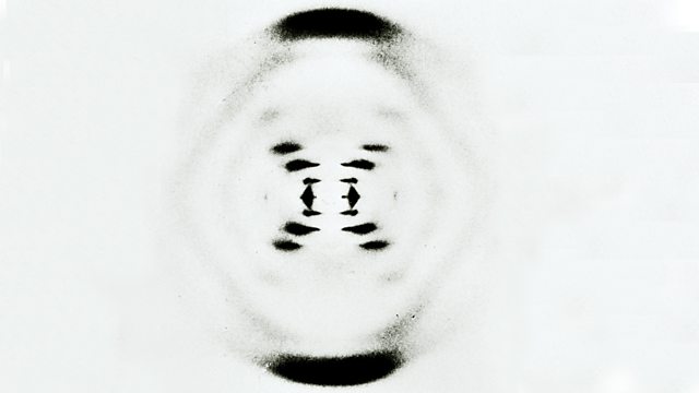
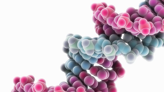
James Watson and Francis Crick worked out the structure of DNA in 1953. By using data from other scientists they were able to build a model of DNA. The X-ray data they used showed that DNA consists of two strands coiled into a double helix.
DNA diffraction
X-ray diffraction photograph of DNA
Synthetic DNA molecule

Base pairs

Each strand of DNA is made of chemicals called bases. Note that these are different to bases in relation to acids and alkalis in chemistry. There are four different bases in DNA:
  • thymine, T
  • adenine, A
  • guanine, G
  • cytosine, C
There are chemical cross-links between the two strands in DNA, formed by pairs of bases. They always pair up in a particular way, called complementary base pairing:
  • thymine pairs with adenine (T–A)
  • guanine pairs with cytosine (G–C)
DNA strand showing bases pairs: TA, GC, AT repeated randomly.
Base pairs on a section of DNA

>